← Back to 4D-STEM explained · 4d User Manual
Why the format matters
A file format is not just a container: it decides what survives of your measurement. Four properties matter for electron microscopy, and the rest of this page is mostly about them.
- Bit depth and number type. Detector counts need 16 bits, reconstructions and phase maps need 32-bit floating point. An 8-bit format (JPEG, GIF) throws away the dynamic range that makes the data quantitative, and cannot represent a negative value at all.
- Lossy or lossless. Lossless formats return exactly the numbers you wrote. JPEG does not: it discards high spatial frequencies — which, in a noisy micrograph or a diffraction pattern, is where the signal lives.
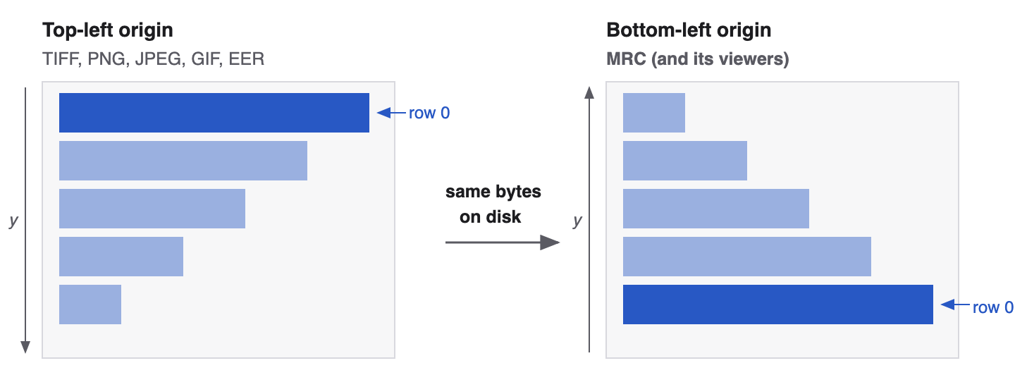
- The origin. Row 0 sits at the top in every ordinary picture format and at the bottom in MRC. Getting this wrong mirrors the image, which flips the handedness of everything derived from it.
- The header. Pixel size, voltage, dose and camera settings only travel with the data if the format has somewhere to put them.
Comparison at a glance
File sizes are for a single 1024 × 1024 image. "Raw" is the pixel data alone; the compressed figures assume noisy detector data, which compresses far less than an ordinary photograph — noise is incompressible, and that is the single most common surprise when people first archive micrographs.
| Format | Compression | Bit depths | Origin | Metadata in the header | 1024 × 1024 |
|---|---|---|---|---|---|
| HDF5 | Optional, lossless, per chunk: gzip, Blosc2, bitshuffle+LZ4, Zstd, LZF | anything: 8/16/32/64-bit integer or float, complex | no convention — whatever the writer used | unlimited, user-defined attributes anywhere in the tree | 2.1 MB at 16 bit raw; counting data often 5–10× smaller compressed |
| MRC | None in the format (files are often gzipped whole) | int8, int16, uint16, float16, float32, complex | bottom-left | fixed 1024-byte header + optional extended header | 2.1 MB at 16 bit, 4.2 MB at 32-bit float, + 1 KB header |
| TIFF | Optional, lossless: LZW, Deflate, PackBits (JPEG-in-TIFF is lossy) | 1–64 bit, integer or float, any number of samples | top-left (Orientation tag 274 can say otherwise) | tags: size, bit depth, resolution, description, date, private tags | 2.1 MB at 16 bit raw; ≈ 1.6–2.0 MB with LZW/Deflate on noisy data |
| EER | Lossless, event-based — the point of the format | event positions, not pixel values | top-left (TIFF container) | TIFF tags plus an XML metadata block | scales with electrons, not pixels — see below |
| PNG | Lossless, Deflate with per-row prediction filters | 8 and 16 bit, grey or RGB(A); no float | top-left | size, bit depth, optional physical pixel size and text chunks | ≈ 1.5–2.0 MB at 16 bit, ≈ 0.7–1.0 MB at 8 bit, on noisy data |
| JPEG | Lossy, DCT-based, quality-dependent | 8 bit per channel | top-left (EXIF Orientation may rotate/flip) | JFIF and EXIF blocks: camera, date, orientation, thumbnail | ≈ 0.2–0.7 MB at quality 90 — noise inflates it |
| GIF | Lossless LZW, but only within a 256-colour palette | 8-bit palette (≤ 256 colours) | top-left | screen descriptor, palette, per-frame delays | ≈ 0.6–1.0 MB after quantising to 256 greys |
Where the origin sits
HDF5 (.h5, .hdf5) — the native format of 4d
HDF5 is not an image format but a hierarchical container: a file holds a tree of groups (like directories), datasets (n-dimensional arrays of any type) and attributes (metadata attached to either). That generality is why it carries a whole 4D-STEM acquisition — scan positions, diffraction patterns, masks, precomputed maps — in one file.
| Compression | Per-chunk filters, all lossless: gzip/Deflate, Blosc2, bitshuffle + LZ4, Zstd, LZF, plus byte-shuffle pre-filters. Chunking also decides read speed: a chunk shaped like one diffraction pattern lets 4d load a single pattern without touching the rest. |
| Bit depth | Anything the machine has, including 64-bit floats and complex numbers. Datasets can be extendable and sparse. |
| Origin | Undefined by the format. An array is just an array; the convention comes from the layout the writer follows. |
| Header | A superblock plus B-trees and heaps, all handled by the library. Scientific metadata lives in user-defined attributes, so the layout must be agreed on — hence the conventions: 4d's own, EMD, NeXus. |
| 1024 × 1024 | Raw size plus a few kilobytes of structure. Compression on counting-mode data, where most pixels are zero, routinely gives 5–10×; on dense 16-bit data expect 1.1–2×. |
4d stores datasets in its version 2.0 convention with Blosc2 compression; the group layout is described in §7 of the main manual.
MRC (.mrc, .mrcs, .map)
The workhorse of cryo-EM: a fixed 1024-byte header, an optional extended header
whose length the fixed header states, then raw pixel data. It stores 2D images, image stacks (.mrcs)
and 3D volumes (.map) in the same structure, which is why a
reconstruction and a particle stack look alike to a reader.
| Compression | None. The format writes raw numbers, which is exactly why it is fast to memory-map and why files are large. Whole files are often gzipped for transfer (.mrc.gz); variants such as MRCZ add Blosc compression but are not part of the standard. |
| Bit depth | Chosen by the mode field: 0 = int8, 1 = int16, 2 = float32, 3 and 4 = complex, 6 = uint16, 12 = float16. Mode 2 is the default for reconstructions, mode 6 for raw camera frames. |
| Origin | Bottom-left by convention — see the figure above. |
| Header | 1024 bytes of fixed fields: image size (nx, ny, nz), mode, start indices, sampling (mx, my, mz), the unit-cell dimensions from which the pixel size follows, axis order, min/max/mean and RMS of the data, space group, size of the extended header, the origin, the MAP stamp, a machine stamp giving the byte order, and up to ten 80-character text labels. An optional extended header follows, which Thermo Fisher (FEI1/FEI2) and SerialEM use for per-frame metadata such as tilt angle, dose and stage position. |
| 1024 × 1024 | 1.05 MB (mode 0), 2.10 MB (modes 1/6), 4.19 MB (mode 2), each plus the 1 KB header. |
TIFF (.tif, .tiff)
TIFF is a tagged container rather than one format: an 8-byte header points to a chain of image file directories, each a list of tags describing one image. That flexibility makes it the usual export format of camera software — and the reason two TIFF files can need completely different code to read.
| Compression | Optional and normally lossless: LZW, Deflate (ZIP), PackBits run-length; newer writers add Zstd and LERC. JPEG compression inside a TIFF is possible and lossy — avoid it for data. On noisy micrographs lossless compression typically saves only 5–20 %. |
| Bit depth | 1 to 64 bits, unsigned, signed or floating point, one or many samples per pixel — one of the few ordinary formats that stores 32-bit float directly. |
| Origin | Top-left by default. Tag 274 (Orientation) can declare any of eight arrangements; many scientific readers ignore it, so a file that relies on it may appear flipped. |
| Header | Byte-order mark (II or MM), magic number 42 (or 43 for BigTIFF), and the offset of the first directory. Tags then give width, height, bits per sample, sample format, compression, strip or tile layout, X/Y resolution and unit. Free-text tags — ImageDescription, Software, DateTime — are where ImageJ, SerialEM and camera vendors hide pixel size, dose and acquisition settings, often as XML or a private tag block. |
| 1024 × 1024 | 1.05 / 2.10 / 4.19 MB raw at 8 / 16 / 32 bit, plus a few hundred bytes of tags; ≈ 1.6–2.0 MB for a noisy 16-bit image with Deflate. |
EER (.eer) — Electron Event Representation
EER takes a different view of what an image is. Instead of integrating the electrons that arrive during a frame into pixel counts, a Falcon 4-class detector records each detected electron as an event: its position, refined to a sub-pixel grid, and the frame in which it arrived. The file is a list of those events, losslessly compressed.
| Compression | Lossless and intrinsic: the gaps between successive events are run-length encoded and entropy coded. Since a low-dose frame is almost entirely empty, this is far more compact than storing mostly-zero pixel arrays — an order of magnitude or more against an equivalent raw movie. |
| Bit depth | Not applicable: there are no pixel values, only event positions. The image appears when you render the events onto a grid, and you choose the grid — the native sensor sampling, or 2× / 4× finer using the sub-pixel positions, and as many or as few frames per rendered image as the dose requires. |
| Origin | Top-left, as the TIFF container implies; the rendering library fixes the convention in practice. |
| Header | EER is wrapped in a TIFF structure, so ordinary TIFF tags describe the frames, with private tags carrying the compressed event bitstreams and an XML block holding the acquisition metadata: sensor size, number of frames, frame rate, dose rate. |
| Size | Proportional to the number of detected electrons, not to the pixel count — roughly one to two bytes per electron. A whole low-dose exposure typically lands in the low hundreds of MB where the equivalent unpacked movie would be several GB. Expressed per 1024 × 1024 area at 1 Å pixels and 40 e−/Ų, the events of the complete exposure amount to a few tens of MB, against 2.1 MB for the single 16-bit integrated image they render into. |
The practical consequence: keep the EER and decide later. Dose weighting, motion correction and the choice of fractionation can all be redone from the events, which is impossible once they have been integrated into fixed frames.
PNG (.png)
| Compression | Lossless Deflate, preceded by a per-row prediction filter (each row is coded as its difference from the row above or the pixel to the left). Excellent on smooth images and line art, poor on noise. |
| Bit depth | 8 or 16 bit, greyscale or colour, with an optional alpha channel. No floating point and no negative values, so a phase map has to be scaled into an integer range before it can be stored. |
| Origin | Top-left; rows are stored top to bottom (optionally interlaced). |
| Header | An 8-byte signature followed by chunks. IHDR carries width, height, bit depth and colour type; pHYs can record the physical pixel size; tEXt/iTXt hold free text. There is no standard place for microscope metadata, so it is usually lost. |
| 1024 × 1024 | ≈ 0.7–1.0 MB for a noisy 8-bit micrograph, ≈ 1.5–2.0 MB at 16 bit; a smooth or largely empty image can drop to a tenth of that. |
PNG is the right choice for figures, screenshots and anything going into a manuscript or a manual — it is lossless, universally readable, and does not invent texture the way JPEG does. It is the wrong choice for data, because the calibration does not travel with it.
JPEG (.jpg, .jpeg)
| Compression | Lossy. The image is divided into 8 × 8 blocks, cosine-transformed, and the high-frequency coefficients are quantised away. At quality 90 the damage is invisible in a photograph; in a micrograph it removes precisely the fine detail you measured, and adds block edges and ringing around strong features. |
| Bit depth | 8 bit per channel. A 16-bit detector image must be scaled down to 256 levels first, which alone rules it out for quantitative work. |
| Origin | Top-left. The EXIF Orientation tag may ask for a rotation or mirror, and viewers disagree about honouring it. |
| Header | Marker segments: JFIF (version, pixel density), optionally EXIF (camera, date, exposure, orientation, GPS, thumbnail), the quantisation and Huffman tables, and the frame header with the dimensions. |
| 1024 × 1024 | ≈ 0.2–0.7 MB at quality 90 for a noisy image — noise defeats the compression, so a micrograph is several times larger than a photograph of the same size. Quality 50 halves it again and the artefacts become obvious. |
GIF (.gif)
| Compression | Lossless LZW — but only after the image has been reduced to a palette of at most 256 colours, which for a micrograph means 256 grey levels. The quantisation, not the compression, is where the data is lost. |
| Bit depth | 8-bit palette indices. One palette entry can be transparent; there is no alpha channel and no 16-bit mode. |
| Origin | Top-left, with each frame positioned by its own left/top offsets inside the logical screen. |
| Header | GIF87a/GIF89a signature, a logical screen descriptor (size, background colour, palette flags), the global colour table, and per-frame descriptors with delay times and disposal methods for animation. |
| 1024 × 1024 | ≈ 0.6–1.0 MB for a noisy greyscale image — LZW can even exceed the raw size on pure noise; a few tens of kilobytes for a plot or a diagram. |
The one thing GIF still does well is a short animation that plays anywhere, such as a tilt series or a through-focus series for a talk. For anything else, PNG is better in every respect.
Other formats you may meet
| DM3 / DM4 | Gatan DigitalMicrograph. A tagged tree carrying the image plus very complete microscope metadata. DM4 uses 64-bit offsets for large files. |
| SER / EMI | Thermo Fisher (formerly FEI) TIA. The .ser holds the data, the companion .emi the metadata — keep the pair together. |
| EMD | Electron Microscopy Datasets: a documented convention inside HDF5, readable by any HDF5 tool. Also the native format of Velox. |
| NeXus | Another HDF5 convention, from the neutron and synchrotron world, increasingly used for electron data. |
| SMV / ADSC | Simple header-plus-data format from X-ray detectors, used by some electron-diffraction pipelines. |
| TVIPS | Movie format of TVIPS cameras, a header followed by concatenated frames. |
| OME-TIFF | TIFF with a standardised XML metadata block in the ImageDescription tag — the light-microscopy answer to the metadata problem. |
Rules of thumb
- Keep the original. Archive what the detector wrote — EER events, the raw movie, the HDF5 — and treat everything else as derived. No conversion adds information.
- Data in HDF5 or MRC, pictures in PNG. HDF5 when the metadata and the structure matter, MRC when the cryo-EM tool chain expects it, PNG for anything a human looks at.
- Never put measurements through JPEG or GIF. Eight bits and lossy compression are not a storage decision, they are a measurement decision — and it is irreversible.
- Check the flip once, deliberately. Convert a deliberately asymmetric image between MRC and TIFF/PNG and look at it. Do it once per pipeline and write down what you found.
- Expect compression to disappoint. Detector noise is incompressible: lossless compression of a dense micrograph saves tens of percent, not factors. Counting-mode and event data are the exception — there the emptiness is real and the savings are large.
- Carry the pixel size. If the format cannot hold it (PNG, JPEG, GIF), record it beside the file, or the image becomes a picture rather than a measurement.